

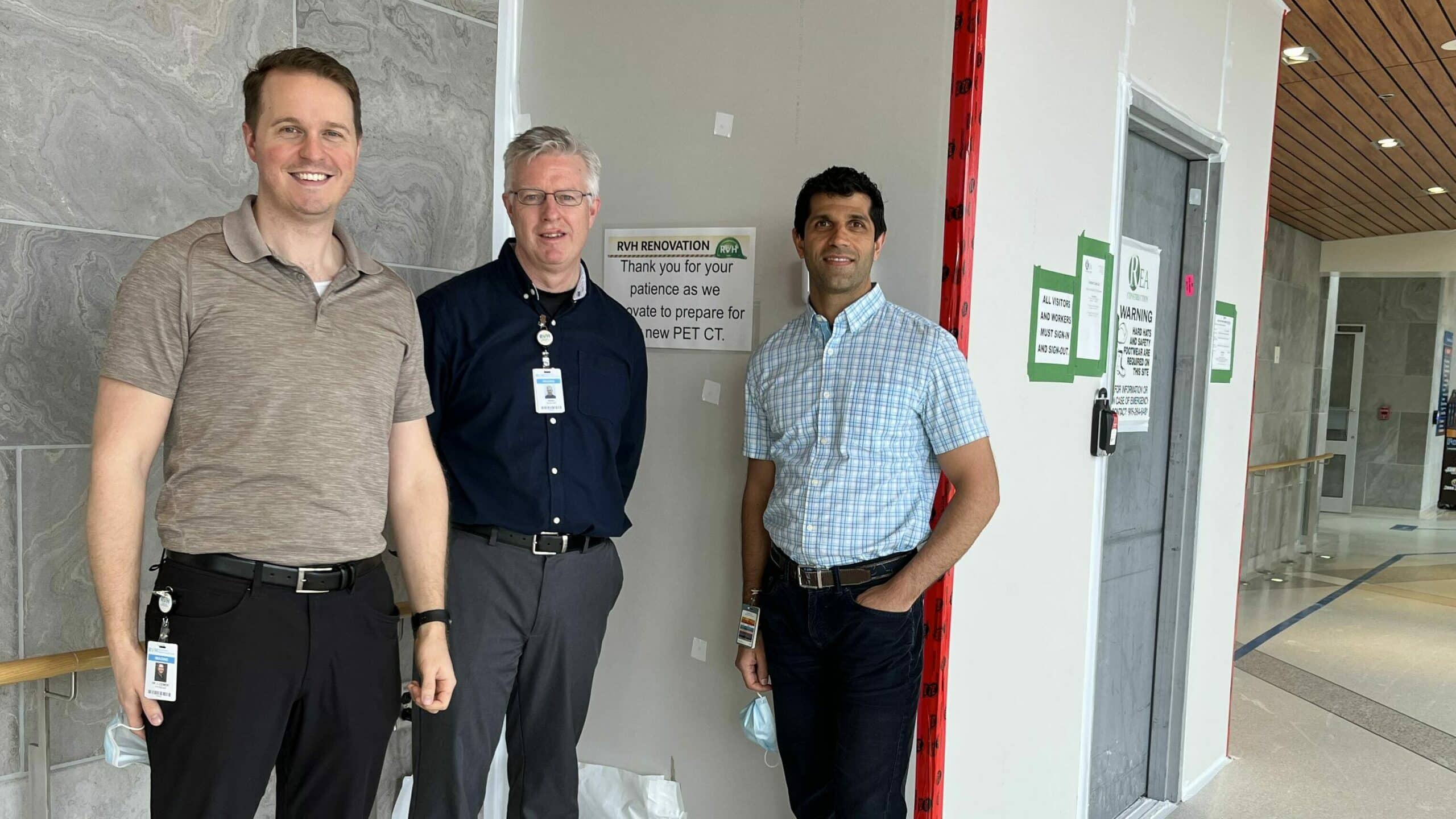
Radiologist and PET-CT Lead, Dr. Cory Ozimok, Manager of Medical Imaging, David Wilson and Medical Director of Imaging, Dr. Raj Grover stand outside the area where construction is underway to make space for the new PET-CT scanner at RVH.
Construction is now underway on the space that will house the new positron emission tomography–computed tomography (PET-CT) scanner at RVH. The PET-CT has arrived and will be installed over the next few months with the goal of it being fully operational and contributing to patient care no later than early fall.
This vital equipment has been funded in part by a generous $500,000 donation from valued RVH donor, Barrie Welding and Machine, as well as support from Jane and Dr. Paul Voorheis, a long-time RVH radiologist, former Imaging Medical Director and interim Chief of Staff. And, also by generous donors like you and through the support of the provincial government.
We asked three of RVH’s leading subject matter experts to give us an overview of the science and to share how this will impact patient care in Simcoe Muskoka. Below is a compilation of answers from radiologist and PET-CT Lead, Dr. Cory Ozimok, Medical Director of Imaging, Dr. Raj Grover, and Manager of Medical Imaging, David Wilson.
How does a PET-CT scanner work?
During a PET scan, patients are injected with a small amount of a radioactive drug called “FDG” or Fluorodeoxyglucose. FDG is a sugar molecule with an additional atom that emits positrons. Cells in the body that are using sugar as energy, including cancer cells, absorb FDG. Because cancer cells require more energy to grow than normal cells, they tend to accumulate more FDG. The PET scanner measures the radiation emitted by the positrons and creates an image of the areas with the highest concentration of FDG. This helps us to detect cancer cells and see tumors based on their metabolic activity. To improve accuracy, PET scanning is often combined with a low dose CT scan, which maps the inside of the body to create a more detailed image.
Most of us have heard of a CT scanner. What makes a PET-CT different?
During a traditional CT scan, radiation is used to create a detailed image of a patient’s body structure. This helps doctors to see the location, size, and shape of tumors. In contrast, during a PET scan, we inject a radiopharmaceutical into the patient and use a scanner to create a functional image of how the body is responding to the radioactive substance. This provides real-time information about the metabolic activity of the tumor. Both CT and PET scans are complementary and provide different information about a tumor, allowing doctors to better understand and treat it.
Who will benefit from us having a PET-CT at RVH?
There are many patients who will benefit from having a PET-CT scanner in our hospital. The majority will be patients from the David and Catherine Hudson Regional Cancer Centre that currently travel great distances to obtain these scans that are crucial for their care. PET-CT is paramount in staging and monitoring patients with lymphoma, lung cancer, esophageal cancer, anal cancer, colorectal cancer, head and neck malignancies, gynecological malignancies, melanoma, multiple myeloma, and breast cancer, among others.
Why do we need this piece of equipment?
PET-CT is now a standard practice for cancer care, and it is becoming more widely used to diagnose and monitor a variety of conditions. Adding a PET-CT scanner to our hospital is a positive step towards improving patient access to this valuable tool. This piece of equipment is essential to provide the highest quality care to patients in Simcoe Muskoka and ensure they receive the latest evidence-based treatments.
What kind of disease or circumstance is a PET-CT used to detect?
In Ontario, PET is primarily used for oncology imaging during a patient’s cancer journey. It helps create a history of the cancer ranging from diagnosis to staging, to monitoring response to treatment and detecting recurrence. There are additional PET radiopharmaceuticals that are highly specific for prostate cancer as well as neuroendocrine cancers that bring with them new targeted therapeutics as well.
Beyond oncology, PET is also useful for cardiovascular disease, including coronary artery disease, myocardial inflammation, vasculitis and in neuroimaging for dementia, Parkinson’s disease, and epilepsy.
How was RVH chosen as a site for the PET-CT?
Currently, Southlake Hospital in Newmarket is in the process of operationalizing a PET-CT as well. They will be up and running around the same time as us. There is no other PET-CT scanner between us and Sudbury.
Provincial PET-CT sites are selected by Ontario Health’s Capital Planning Advisory Committee using data to evaluate criteria such as geographic access for patients and that the demand for scans is enough to ensure economic viability (ideally at least 700 to 1,000 scans per year at a minimum).
PET-CT cannot operate without a cyclotron supply of isotopes. Choosing a location for a PET-CT must also factor in the distance from the nearest cyclotron facility and the feasibility of shipping to the site.
What is involved in getting our space ready for the PET-CT in our Medical Imaging Department?
To make the space fit for use, we’re constructing rooms for the scanner, patient preparation, and patient holding and resting prior to having their scan. Everything must be lead-lined to ensure radiation safety. The floor also needs to be able to hold the weight of the combined PET and CT scanners. We have architects and a physicist on contract who are assisting us with room design, layout, radiation safety and necessary ministry approvals.
What does a PET-CT cost?
Like a car, there are different makes, models, trims, and accessories. The Siemens Vision 600 scanner we are getting is approximately $2.4M for the base model, but with the additional features we require, our total to closer to $3.3M.
Will the PET-CT be operational 24/7?
It will ramp up as demand increases. The radioisotopes that make the radioactive injectable a patient needs for the scan to work only have a short half-life (minutes to hours). To make the most of those radiopharmaceuticals, PET scanners generally only run during daytime hours. We will be getting our radioisotopes from a company named Isologic that distributes isotopes from a cyclotron based at Sunnybrook Hospital in Toronto.
Will the PET/CT have phased uses?
Yes, the primary area of focus here will start with oncology patients and the radioactive pharmaceutical called FDG (fluorodeoxyglucose) which has wide use in PET cancer scanning. FDG is the most common PET tracer used. Once we get up to speed with oncology scanning, we will look at the feasibility of doing some cardiac PET scans and then possibly some neurological scans.
Will this require additional staff to operate?
The scanner is being installed as part of RVH’s Nuclear Medicine department, but because we are adding an entirely new scanner, we need additional staff to operate it.
We have already hired one part-time technologist and are in the process of hiring a second. We have put a senior PET-CT technologist in place whom we sponsored to train and get a specialty certificate in this area. Two of our current nuclear medicine techs are working on their certification.
From a radiologist perspective, Dr. Ozimok has done training in PET-CT and the RVH group is looking to add a second PET-CT-trained radiologist. More technologist staff will need to be added as the scanner ramps up to 5 days/week.
Who performs the scans? Is it one person, a team?
One technologist can do all aspects of patient prep and scanning, but once more than four patients are booked per day, a team is required to keep the flow going. The half life of FDG is short and the scan time is about 15-20 minutes, so to ensure the timing is perfect even on a busy scan day, two or three technologists are needed to keep things moving.
What is the experience like for a patient?
Patients are in the department for about two hours from start to finish, even though the scan time is only a fraction of that. They need to complete a pre-scan questionnaire, have an IV injection of FDG, wait quietly for one hour, and then have the scan.
Donate today to help us bring more leading-edge technology, like the PET-CT scanner, to RVH. Your generous gift has the power to make a lasting impact on the lives of countless individuals and will help us advance medical care and improve health outcomes for our community.